Anatomy Of Chest Area - Images 04. Skeletal System | Basic Human Anatomy : Tuhansia uusia ja laadukkaita kuvia joka päivä.. The chest anatomy includes the pectoralis major pectoralis minor and the serratus anterior. Less frequently areas of decreased density are seen as in emphysema or lungcysts. Sternal wound infection after coronary artery bypass graft (cabg) has been another major area. Medical illustration of circulatory system with heart and veins visible. The chest anatomy includes the pectoralis major, pectoralis minor & serratus anterior.
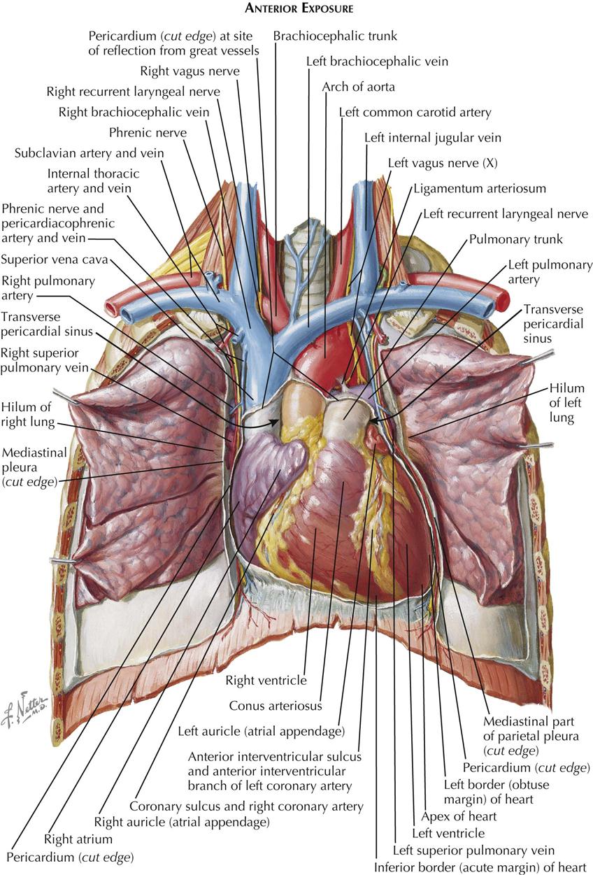
It is where the left ventricle hits against the chest wall. Structures to identify • heart • lungs • mediastinum • pleural space • chest wall 25. Pathology of the heart, mediastinum, lungs and pleura. Is its effect so thoroughly nebulous that it's hard to justify? In insects, crustaceans, and the extinct trilobites, the thorax is one of the three main divisions of the creature's body.
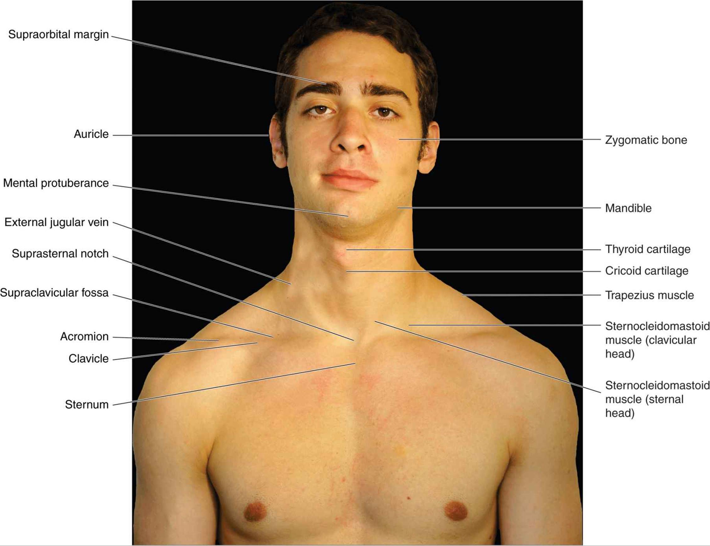
1. Anatomy | Thoracic Key from thoracickey.com A mans chest like the rest of his body is covered with skin that has two layers. Pertinent anatomy demonstrated the chest radiograph should demonstrate all of the anatomy of the lungs from the apices to the lung bases. Diagrams of normal venous anatomy of the thorax. Is the book of chest anatomy almost entirely pointless? Structures that pass through this area can be thought of as the birds of the mediastinum: The chest is the area of origin for many of the body's systems as it houses organs such as the heart, esophagus, trachea, lungs, and thoracic diaphragm. Book of chest anatomy is a passive item. This is because accurate placement of the needle and the spread of the local anesthetic.
Is the book of chest anatomy almost entirely pointless?
Pertinent anatomy demonstrated the chest radiograph should demonstrate all of the anatomy of the lungs from the apices to the lung bases. Anatomy of the chest and the lungs: These lungpatterns will discussed in more detail in an article. • a chest mri may be done for the following. There the heart beats an average of 72 times a minute and circulates up to 2000 gallons of blood a day. The circulatory system does most of its work inside the chest. Iv contrast may be injected into a vein in the patient's arm or hand. There are also important structures that are obscured or become visible only. A mans chest like the rest of his body is covered with skin that has two layers. See more of our training videos here: Understand the different muscle groups that make up the chest, as part of our new series called muscle anatomy. Reading of chest radiographs some basic anatomy and physiology; However, once the anatomic layers and tissue sheets are dissected, the anatomy of nerve structures without the tissue sheaths around them is of little relevance to the clinical practice of regional anesthesia.
Related posts of anatomy of the chest area. Profile view of female chest area. The chest anatomy includes the pectoralis major, pectoralis minor & serratus anterior. Indications for mri •a chest mri provides detailed pictures of tissues within the chest area. Ct anatomy of the chest, axial reconstruction.
Pectoral anatomy from www.edoctoronline.com Diagrams of normal venous anatomy of the thorax. Anatomy of the chest and the lungs: Structures that pass through this area can be thought of as the birds of the mediastinum: These lungpatterns will discussed in more detail in an article. Understand the different muscle groups that make up the chest, as part of our new series called muscle anatomy. Huge collection, amazing choice, 100+ million high quality, affordable rf and rm images. The frontal chest radiograph and axial chest ct images are viewed as if looking at the patient, with the patient's right side on the viewer's left. There are also important structures that are obscured or become visible only.
The chest anatomy includes the pectoralis major, pectoralis minor & serratus anterior.
Pathology of the heart, mediastinum, lungs and pleura. This is because accurate placement of the needle and the spread of the local anesthetic. Is the book of chest anatomy almost entirely pointless? Each of these anatomical structures should be viewed using a systematic approach. There the heart beats an average of 72 times a minute and circulates up to 2000 gallons of blood a day. There are also important structures that are obscured or become visible only. You can observe for it and. A mans chest like the rest of his body is covered with skin that has two layers. Is its one synergy actually worthwhile? Removed from the chest area is appropriate. Related posts of anatomy of the chest area. Structures that pass through this area can be thought of as the birds of the mediastinum: The circulatory system does most of its work inside the chest.
The chest anatomy includes the pectoralis major pectoralis minor and the serratus anterior. It is where the left ventricle hits against the chest wall. Is the book of chest anatomy almost entirely pointless? Improves the contents of broken chests. Structures that pass through this area can be thought of as the birds of the mediastinum:
Atlas of Surface Anatomy - Hadzic's Peripheral Nerve ... from doctorlib.info Venous circulation of the bronchia into the azygos and hemiazygos veins. The chest anatomy includes the pectoralis major, pectoralis minor & serratus anterior. Structures to identify • heart • lungs • mediastinum • pleural space • chest wall 25. Understand the different muscle groups that make up the chest, as part of our new series called muscle anatomy. Is its one synergy actually worthwhile? The chest anatomy includes the pectoralis major pectoralis minor and the serratus anterior. Reading of chest radiographs some basic anatomy and physiology; Book of chest anatomy is a passive item.
Learn about each muscle, their locations & functional anatomy.
Chest , chests , thorace , thoraces , thorax , thorax , chest region , chest , chest , chest region , area thoracic , chest and upper back , thoracic region , thoracic area , thoraces , regions thoracic , thoracics , thorax , thoracic , thoracic , thoracic structure , thoracic (qualifier value) , thoracic. Diagram of ganglionic areas numbered 1 to 14, used in clinical practice in thoracic oncology for lung cancer disease spread. Is its effect so thoroughly nebulous that it's hard to justify? It is where the left ventricle hits against the chest wall. Pertinent anatomy demonstrated the chest radiograph should demonstrate all of the anatomy of the lungs from the apices to the lung bases. Iv contrast may be injected into a vein in the patient's arm or hand. Tuhansia uusia ja laadukkaita kuvia joka päivä. Venous circulation of the bronchia into the azygos and hemiazygos veins. Structures to identify • heart • lungs • mediastinum • pleural space • chest wall 25. Is its one synergy actually worthwhile? Is the book of chest anatomy almost entirely pointless? Learn about chest anatomy with free interactive flashcards. Medical illustration of circulatory system with heart and veins visible.
Is the study of human anatomy complete or has it gone nano? answered by dr anatomy of chest. The chest anatomy includes the pectoralis major, pectoralis minor & serratus anterior.

Belum ada Komentar untuk "Anatomy Of Chest Area - Images 04. Skeletal System | Basic Human Anatomy : Tuhansia uusia ja laadukkaita kuvia joka päivä."
Posting Komentar